

Minimally Invasive & Open Heart
Understanding Atrial Septal Defect
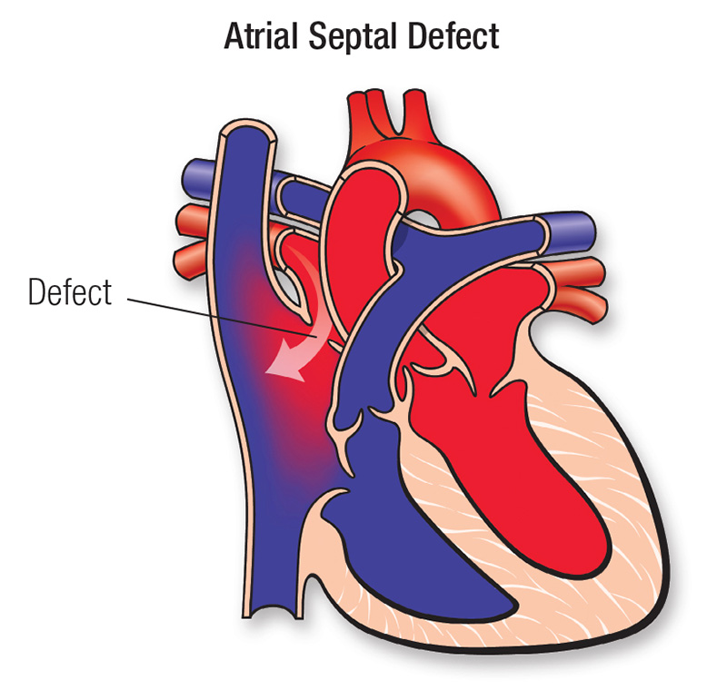
Atrial Septal Defect (ASD) is a congenital heart defect where there is an abnormal opening in the wall (septum) between the two upper chambers of the heart (atria). This opening allows oxygen-rich blood to mix with oxygen-poor blood, which can increase the workload on the heart and lungs over time. Small ASDs may not cause symptoms, but larger defects can lead to serious complications if left untreated.
- Shortness of breath, especially during activity
- Fatigue and reduced exercise capacity
- Frequent respiratory infections
- Heart palpitations or irregular heartbeat
- Swelling in legs, feet, or abdomen (in advanced cases)
- Heart murmur (detected during examination)
- Stroke (rare, in untreated cases due to clot passage)
- Electrocardiogram (ECG)
- Echocardiogram (2D/3D Echo with color Doppler)
- Chest X-ray
- Transesophageal echocardiography (TEE)
- Cardiac MRI (in selected cases)
- Oxygen saturation studies
Device Closure (Minimally Invasive Treatment)
A catheter-based procedure where a closure device is inserted through a blood vessel and placed to seal the hole in the heart. This is the most common treatment for suitable ASDs.
Open Heart Surgery
Recommended when the defect is large, not suitable for device closure, or associated with other heart abnormalities. The hole is directly closed using sutures or a patch.
Medical Management
Used only for symptom control or complications such as heart failure or arrhythmia; it does not close the defect.
- No large chest incision
- Short hospital stay
- Less pain and faster recovery
- Minimal scarring
- Quick return to normal activities
- Large or complex ASD not suitable for device closure
- Deficient septal rims preventing device placement
- Associated congenital heart defects requiring correction
- Failed previous device closure attempt